
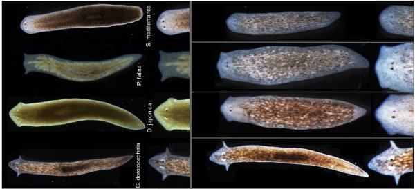
In a finding that challenges long-held assumptions about how an organism's body plan is determined, biologists at Tufts University have coaxed a species of flatworm to grow heads and brains characteristic of a different species—without modifying its genetic code. The research, published as the cover story in the November 2015 issue of the International Journal of Molecular Sciences, demonstrates that electrical communication between cells can override a species' typical anatomy.
The experiments centered on Girardia dorotocephala, a free-living planarian known for its extraordinary regenerative abilities. By interrupting the electrical signals that pass between cells, the team triggered the worms to regenerate heads and brains resembling those of other planarian species. Notably, the altered anatomy extended beyond the head shape: the distribution of adult stem cells also shifted, indicating that the bioelectric changes influenced multiple aspects of development.
This work adds to a growing body of evidence suggesting that an organism's form is not strictly dictated by its DNA sequence. While chromosomes have long been viewed as the blueprint for anatomy, the Tufts findings indicate that information outside the genome—specifically, the electrical synapses between cells—can play a decisive role in shaping body structure. The researchers note that the effect was temporary; after a few weeks, the worms reverted to their own species' head shape, underscoring the need for further investigation.
Implications for Regenerative Medicine
The study's authors believe the results could eventually inform treatments for birth defects and techniques for regenerating damaged or missing body parts in humans. However, they acknowledge that such applications remain distant, as the mechanisms involved are not yet fully understood. The ability to influence regeneration through bioelectric signals, rather than genetic editing, opens a new avenue for therapeutic exploration.
Maya Emmons-Bell, a Tufts undergraduate senior majoring in biology, served as the paper's first author. In a university press release, she highlighted the significance of the work: “We’ve demonstrated that the electrical connections between cells provide important information for species-specific patterning of the head during regeneration in planarian flatworms,” she said. “This kind of information will be crucial for advances in regenerative medicine, as well as a better understanding of evolutionary biology.”
The research was conducted at Tufts, where scientists have long studied planarian regeneration as a model for understanding tissue repair. The current study builds on that foundation, suggesting that bioelectric signals are a key factor in determining how regenerating tissues are patterned.
While the prospect of using such techniques in humans is speculative at this stage, the findings offer a proof of concept that anatomy can be influenced without altering the genome. The team plans to continue investigating how these electrical signals are generated and how they interact with genetic pathways, with the hope of one day translating the insights into clinical applications.
[IMG_1]
For now, the study stands as a reminder that the rules of biology are not as fixed as once thought. As Emmons-Bell noted, the work provides critical information for both regenerative medicine and evolutionary biology, and it demonstrates that even an undergraduate can contribute meaningfully to cutting-edge science.
Comments 0